W.B.C.S. Examination Notes On – Diaphragm Anatomy – Medical Science Notes.
The diaphragm is an unpaired, dome shaped skeletal muscle that is located in the trunk. It separates the thoracic and abdominal cavities from each other by closing the inferior thoracic aperture.Continue Reading W.B.C.S. Examination Notes On – Diaphragm Anatomy – Medical Science Notes.
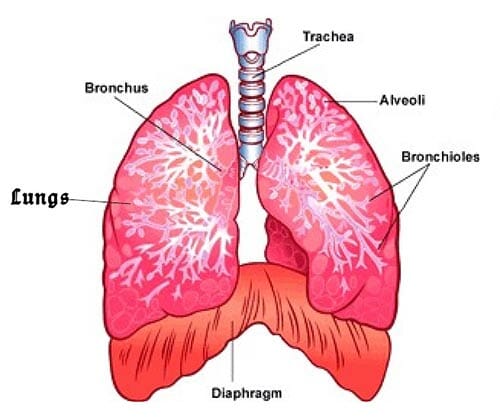
The diaphragm is the primary muscle that is active in inspiration. Contraction of the muscle facilitates expansion of the thoracic cavity. This increases volume of the the cavity, which in turn decreases the intrathoracic pressure allowing the lungs to expand and inspiration to occur.
The diaphragm is much more than just a sheath separating your thoracic and abdominal cavities. This article will examine this intricate and crucial muscle in detail, looking at its anatomy, function and structures which pass through it.
| Origin | Sternal part: Posterior aspect of xiphoid process
Costal part: Internal surfaces of lower costal cartilages and ribs 7-12 Lumbar part: Medial and lateral arcuate ligaments (lumbocostal arches), bodies of vertebrae L1-L3 (+intervertebral discs), anterior longitudinal ligament |
| Insertion | Central tendon of diaphragm |
| Relations | Pleural cavities, pericardial sac, liver, right kidney, right suprarenal gland, stomach, spleen, left kidney, left suprarenal gland |
| Openings | Aortic hiatus (aorta, azygos vein, thoracic duct), esophageal hiatus (esophagus, vagus nerve), caval hiatus (inferior vena cava)
Greater, lesser, least splanchnic nerves, superior epigastric vessels |
| Innervation | Phrenic nerves (C3-C5) (sensory innervation of peripheries via 6th-11th intercostal nerves) |
| Blood supply | Subcostal and lowest 5 intercostal arteries, inferior phrenic arteries, superior phrenic arteries |
| Functions | Depresses costal cartilages, primary muscle of breathing (inspiration) |
Structure and relations
The diaphragm is a musculotendinous sheet. It has three muscular parts (sternal, costal, and lumbar), each have their own origin and all insert into the central tendon of diaphragm. The diaphragm is shaped as two domes, with the right dome positioned slightly higher than the left because of the liver.
The diaphragm has two surfaces: thoracic and abdominal. The thoracic diaphragm is in direct contact with the lungs and pericardium, while the abdominal diaphragm is in direct contact with the liver, stomach, and spleen.
Anatomically, you can define hiatus as an opening, slit, or gap that allows structures to pass. These openings in the diaphragm allow the inferior vena cava, esophagus, vagus nerves, descending aorta, and other structures to pass through.
The muscles of the diaphragm arise from the lower part of the sternum (breastbone), the lower six ribs, and the lumbar (loin) vertebrae of the spine and are attached to a central membranous tendon. Contraction of the diaphragm increases the internal height of the thoracic cavity, thus lowering its internal pressure and causing inspiration of air. Relaxation of the diaphragm and the natural elasticity of lung tissue and the thoracic cage produce expiration.
The diaphragm is also important in expulsive actions—e.g., coughing, sneezing, vomiting, crying, and expelling feces, urine, and, in parturition, the fetus. The diaphragm is pierced by many structures, notably the esophagus, aorta, and inferior vena cava, and is occasionally subject to herniation (rupture). Small holes in the membranous portion of the diaphragm sometimes allow abnormal accumulations of fluid or air to move from the abdominal cavity (where pressure is positive during inspiration) into the pleural spaces of the chest (where pressure is negative during inspiration). Spasmodic inspiratory movement of the diaphragm produces the characteristic sound known as hiccupping.
Please subscribe here to get all future updates on this post/page/category/website


 Toll Free 1800 572 9282
  mailus@wbcsmadeeasy.in


